


Key Points
- Very significant time savings over traditional methods
- Clear, easy to interpret results
- Simple, rapid operation allows use at point-of-care
 For this overview the term ‘enteric viruses’ refers to an important, but diverse, group of viruses found in the intestinal tract of humans and animals. Some of the most important are listed in table 1 below. They are mainly associated with diarrhoea and gastroenteritis, usually mild and self-limiting, although some members of the group cause asymptomatic infections.
Enteric viruses are the commonest causes of gastroenteritis worldwide, they are most often transmitted via the faecal-oral route, with transmission by direct human contact and via fomites being common. The infective dose can be very low. For example, a single rotavirus is capable of causing human infection. Some enteric viruses, notably noroviruses, are highly infectious and may be easily spread in aerosols and by contact with contaminated surfaces, sometimes resulting in large outbreaks of illness.
Enteric viruses may also be present in contaminated water supplies and waterborne outbreaks of disease are not uncommon. Foodborne transmission has been shown to occur in some outbreaks of viral gastroenteritis.
| Table 1. Some Clinically Significant Enteric Viruses | ||
| Family | Genus | Types |
| Astroviridae | Mamastrovirus | human astroviruses |
| Adenoviridae | Mastadenovirus | human adenoviruses |
| Caliciviridae | Norovirus | noroviruses (Norwalk-like viruses) |
| Sapovirus | human sapoviruses | |
| Parvoviridae | Parvovirus | human parvoviruses |
| Picornaviridae | Enterovirus | Non-polio enteroviruses: cocksackievirus A & B, echoviruses, human enteroviruses (types 68 to 71) |
| Reoviridae | Rotavirus | human rotaviruses |
Detection and isolation techniques
Sampling and Preparation Appropriate sampling procedures are critical for the successful isolation and detection of enteric viruses. Materials collected may include clinical samples, such as stools, and non-clinical samples, such as water and foods.
Water samples are tested both for specific viral contamination and for indicators of faecal contamination, such as adenoviruses and enteroviruses. Since viruses are only likely to be present in water at low levels, very large samples of many litres may need to be collected. Testing water samples for enteric viruses requires a specialised concentration step prior to isolation and detection. Methods used with some success include adsorption-elution techniques, precipitation and ultrafiltration.
Testing of food samples for enteric viruses is not routinely practiced, other than for specific commodities, particularly bivalve molluscs, which concentrate viral particles within their bodies when feeding in contaminated water and are a known hazard for enteric virus infection. Specific sampling and virus concentration methods have been developed for enteric virus detection in shellfish.
Clinical samples, especially stool samples, are the main application for routine testing for enteric viruses. There are established procedures for taking clinical samples for diagnostic virus testing and these should be closely followed to maximise the chances of detecting the causative organism. Some enteric viruses are quite resistant to freezing and samples can be stored at –20oC if longer term storage is required.
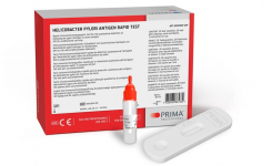
Immunological Detection Methods The majority of immunological methods for enteric viruses are based on antigen detection and employ enzyme immunoassay (EIA), latex agglutination, or immunochromatography technologies.
A wide range of commercial products has been developed and many of these are available as easy-to-use kits that can be applied at the point-of-care. For example, a number of slide-based latex agglutination kits and lateral flow immunochromatography test strips are available for the detection of rotavirus and adenoviruses in suspended stool samples.
These tests offer simplicity in operation and can be performed outside of the laboratory to provide a rapid diagnosis. Their main disadvantages are that they are usually only qualitative tests, have limited sensitivity and cannot distinguish infective viruses.
Molecular Detection Methods Most enteric viruses, apart from adenoviruses and parvoviruses, are RNA viruses, and most molecular techniques therefore rely on detecting specific viral RNA sequences. By focusing on the viral genome, molecular methods can improve specificity and sensitivity significantly. A number of nucleic acid hybridisation methods have been developed for enteric viruses, including dot-blot and sandwich hybridisation techniques, but their sensitivity is often little better than that of conventional techniques.
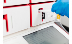
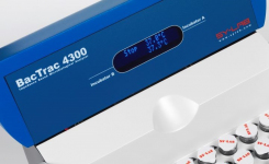
Techniques that allow the amplification of target nucleic acid sequences have revolutionised enteric virus detection in clinical and non-clinical samples. The principal amplification technique used for RNA viruses is reverse-transcriptase polymerase-chain reaction (RT-PCR). Theoretically, this technique is capable of detecting a single virus within 24 hours. Detection of the amplified sequence may be done at the reaction endpoint, or by continuous monitoring (real-time PCR). Real-time PCR has the advantage of allowing the virus to be quantified. A number of commercial applications are available for detection of enteric viruses, particularly enteroviruses. Multiplex PCR methods have been developed that allow the detection of more than one nucleic acid sequence, and therefore more than one virus type, in the same assay.
Other nucleic acid amplification methods have also been developed, notably nucleic acid sequence base amplification (NASBA). This technique is also known as isothermal amplification, as it does not require the repeated temperature cycling used in conventional PCR methods. At least one commercial application of NASBA has been developed for enterovirus detection. The advantages of PCR and related methods are considerable. The main benefit is time saving – 24 hours to a result rather than several days or weeks for a cell culture assay. PCR also requires less operator skill and training to carry out and can be automated to process large numbers of samples. It is extremely sensitive, although it can be vulnerable to contamination and cannot distinguish infective viruses. Most PCR methods also require some costly equipment and are not suitable for use outside the laboratory. Non-quantitative PCR results may be difficult to interpret, since low numbers of virus that do not signify an infection may be detected, but this problem is largely overcome by real-time PCR.
Get the latest updates in Rapid Microbiological Test Methods sent to your email? Subscribe to the free rapidmicrobiology eNewsletter