


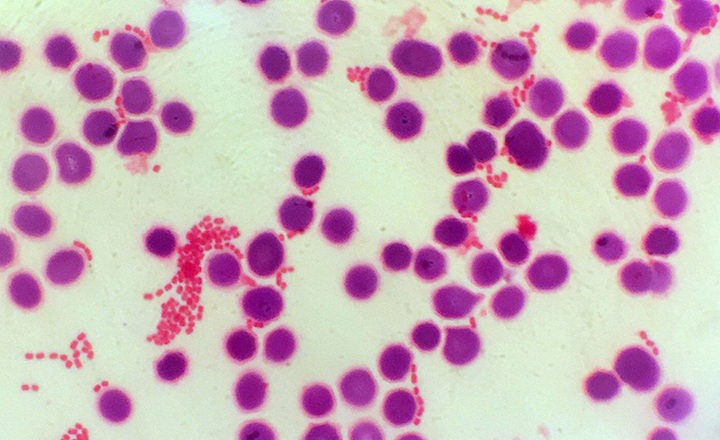
- Gram stain interpretation from positive blood cultures remains one of the most time-sensitive and operator-dependent steps in diagnosing bloodstream infections, directly impacting early antimicrobial therapy decisions.
- Delays or variability in interpretation can affect patient outcomes, as each delay in appropriate therapy increases mortality risk in bloodstream infections.
- Traditional microscopy is labor-intensive and subjective, requiring skilled personnel and limiting scalability in high-throughput or resource-limited settings.
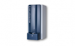
- Emerging imaging modalities such as digital holographic microscopy (DHM) provide label-free, quantitative imaging and enable computational reconstruction of specimens, making them well-suited for integration with AI-driven analysis.
Key Findings: This study by Smith et al. integrates digital holographic microscopy with AI-based image analysis to interpret Gram stains from positive blood cultures versus interpretation performed by clinical scientists.1 Holograms were collected from a total of 532 slides.
- Accuracy: On validation using slides not included in training, the network achieved 99% accuracy for Gram classification compared with expert interpretation.
- AI-driven classification of organisms: Machine learning models were trained to identify Gram morphology and bacterial categories. Automated accuracy varied by morphology:
- 93.3% for Gram-negative bacilli
- 95.2% for Gram-positive cocci in clusters
- 75% for Gram-positive cocci in chains
- Reduction in manual workload: enables automated prescreening and classification, reducing reliance on technologist-driven microscopy and potentially improving laboratory throughput.
- Digitization enables remote and standardized review: Digital output allows image storage, remote interpretation, and auditability.
Bigger Picture: This study highlights the ongoing shift toward AI-enabled, fully digitized clinical microbiology workflows, where traditionally manual tasks are increasingly automated. Gram stain interpretation from positive blood cultures remains a time-critical yet operator-dependent step, and automation using digital holographic microscopy offers a path toward faster and more standardized results.
The ability to generate digital, quantitative images enables consistent interpretation, remote review, and improved documentation, addressing long-standing variability associated with manual microscopy. These capabilities also support scalability in high-volume laboratories and may help mitigate workforce shortages affecting clinical microbiology.
As with other AI-driven diagnostic tools, widespread adoption will depend on robust validation across diverse organisms and specimen types, seamless workflow integration, and regulatory acceptance. Overall, this work reflects a broader movement toward data-driven, automated microbiology laboratories, where imaging and interpretation are increasingly unified into integrated diagnostic systems.
(Image Credit: iStock/Scharvik)