For information on COVID-19 please go to Detection of SARS-CoV-2 Causative Agent of COVID-19
Key Points
- Significant time savings over traditional methods
- Screening kits allow detection of several different viruses in a single assay
- Simple, rapid operation allows use at the point-of-care
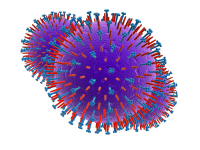 The respiratory viruses are a large and diverse group that includes some of the commonest causes of human viral infection worldwide. They are capable of causing a wide range of diseases, from mild self-limiting upper respiratory tract infections, such as sore throats and the common cold, to more serious infections like influenza, bronchiolitis, potentially life threatening pneumonia and severe acute respiratory syndrome (SARS). Some of the most important types from a clinical point of view are shown in table 1 below.
Historically, some respiratory viruses, notably the influenza viruses, have been associated with highly destructive global pandemics causing millions of deaths. For example, the so-called ‘Spanish flu’ pandemic of 1918-19 is estimated to have caused many more deaths than the Great War of the preceding four years – probably at least 50 million worldwide. There are serious current concerns that the emergence of new influenza virus types in the human population, through mutation or by antigenic shift between avian and human viruses, could lead to further pandemics in the future.
The respiratory viruses are most often transmitted via the aerosol route by inhaling infected droplets, although mechanical transmission through the nose or eye following direct contact with an infected person may also play a significant role in some cases. Infected individuals usually produce large numbers of infective particles in their respiratory secretions, with a peak often seen soon after the onset of symptoms. Many respiratory virus infections, especially those caused by respiratory syncytial virus (RSV) and the common cold viruses, are highly contagious and will spread rapidly through a population, especially in crowded conditions.
Respiratory virus infections are extremely common worldwide. For example, acute viral nasopharyngitis – more often known as the common cold – is the most common type of human infection and children may have a many as six to eight episodes every year. RSV is the commonest cause of lower respiratory tract infection in young children and influenza viruses are also major causes of human disease carrying significant costs in terms of health care and lost economic productivity. Pandemic respiratory infections have the potential to be among the most serious threats to global human health posed by any biological agent.
| Table 1. Some Clinically Significant Respiratory Viruses | |||
| Family | Genus | Types | Diseases caused |
|---|---|---|---|
| Adenoviridae | Mastadenovirus | Human adenoviruses | Pharyngitis, pneumonia, gastroenteritis, conjunctivitis |
| Coronaviridae | Coronavirus | Human coronaviruses, SARS coronavirus | Common colds, severe acute respiratory syndrome (SARS) |
| Orthomyxoviridae | Influenzavirus A | Influenza A, Avian influenza A | Influenza, bronchiolitis, pneumonia, severe lower respiratory tract infection |
| Influenzavirus B | Influenza B | Influenza | |
| Paramyxoviridae | Metapneumovirus | Human metapneumovirus | Pharyngitis, bronchiolitis, pneumonia |
| Pneumovirus | Human respiratory syncytial virus (RSV) | Croup, bronchiolitis, pneumonia (in infants and young children) | |
| Respirovirus | Human parainfluenza virus types 1 & 3 | Croup, non-specific upper respiratory tract infections, bronchopneumonia (mainly in children) | |
| Rubulavirus | Human parainfluenza virus types 2 & 4 | Croup, pharyngitis and colds (mainly in children) | |
| Parvoviridae | Bocavirus | Human bocaviruses | Thought to be associated with a variety of respiratory infections, including bronchiolitis and pneumonia |
| Picornaviridae | Rhinovirus | Human rhinovirus A & B | Common colds |
Detection and isolation techniques
Diagnostic tests for viruses in general fall into one of three categories.
1. Direct examination of the sample – electron microscopy, antigen detection, molecular detection of viral genomes 2. Indirect testing – cell culture techniques and animal tests 3. Serology – detection of specific serum antibodies
Methods for the isolation and detection of respiratory viruses in all three categories have been developed, but the preferred method depends on the individual virus. However, sampling and specimen collection are always an important factor.
Sampling Appropriate sampling procedures are critical for the successful isolation and detection of respiratory viruses. Only clinical samples are routinely collected, and these may include nasal and throat swabs, nasopharyngeal aspirates, tracheal aspirates, broncho-alveolar lavage fluid and serum. There are established procedures for taking clinical samples for diagnostic virus testing and these should be closely followed to maximise the chances of detecting the causative organism. For example, swabs should be transported in viral transport medium and all samples should be refrigerated if there is a delay of more than 2 hours in reaching the laboratory.
Traditional detection and isolation methods The principal traditional or classical techniques for detecting and isolating respiratory viruses are indirect cell culture methods and direct electron microscopy. Most respiratory viruses can be detected by culture in common cell lines specific to the virus concerned. This allows both quantification and isolation of the virus. Cell lines used include green monkey kidney, Hep-2 and Madin-Darby canine kidney (MDCK) cells. Traditionally, cell line cultures are inoculated with the sample and then incubated for up to a week, before examination for any evidence of cytopathogenic effects (CPE), such as damaged cells, or the sloughing off of the cell monolayer. Positive and negative results are then confirmed by further passages (subculturing) in the specific cell line. Unfortunately, some respiratory viruses, notably the influenza viruses, produce little or no CPE and confirmation and identification of positive cell cultures was normally done using haemadsorption, or haemagglutination tests. More recently however, immunofluorescence, or molecular tests have been used for confirmation and identification. This improves the sensitivity and specificity of the test and gives a significant time saving.
Modified culture methods allowing more rapid detection are commonly used for isolation of respiratory viruses today. Foremost among these methods is shell vial culture, in which the clinical specimen is inoculated onto a cell monolayer that has been grown on a cover slip in a shell vial culture tube. After inoculation the tube is centrifuged at low speed and then incubated. The centrifugation process is thought to make the cells more susceptible to viral infection and thus reduce the detection time. Shell vial culture is used to identify adenoviruses, influenza A & B, parainfluenza viruses and RSV and can achieve time savings of several days, especially when used with immunofluorescence, or molecular confirmation methods. Shell vial cultures containing mixed cells lines, such as R-Mix cells, can also be used as a rapid screening test for respiratory viruses. The technique can reliably detect more than one virus type if combined with several different specific fluorescent antibodies.
Cell culture methods have some major drawbacks. They are often time consuming – although this problem has been overcome to some extent by modified culture techniques – and require highly trained staff in specialised laboratories. Interpretation of results can also be difficult and requires experienced staff. On the other hand, cell culture is relatively sensitive, will detect virus infectivity and allows the result to be quantified.
Electron microscopy (EM) is now little used for the detection of respiratory viruses in samples or in culture. However, some types, such as parainfluenza viruses, the SARS virus and adenoviruses, have a characteristic appearance and can be presumptively identified by direct examination, using a negative staining technique, or by immuno-electron microscopy, where a specific antibody is used to agglutinate, or to capture virus particles in the sample. EM depends on there being a large number of virus particles (at least 105-106 per gram) present in the sample.
Rapid methods In general, rapid methods for the detection of respiratory viruses are based either on immunological, or on molecular techniques.
Immunological methods The majority of immunological methods for respiratory viruses are based on antigen detection and employ immunofluorescence, enzyme immunoassay (EIA), or immunochromatography technologies. Direct (DFA) and indirect immunofluorescence (IFA) methods are widely used for detection of respiratory viruses. DFA uses a specific labelled antibody to probe for a viral antigen, while IFA uses an unlabelled antibody, followed by a labelled antibody specific to the first probe – allowing some amplification and improved sensitivity. Detection is performed using fluorescence microscopy. Both DFA and IFA methods are quite labour intensive and need highly trained staff, but are very useful techniques for confirming and identifying viruses in clinical samples and in positive cell cultures. A number of commercial products are available, including screening kits for the detection of up to seven different respiratory viruses in culture.
Commercial immunoassay-based products have also been developed as easy-to-use kits that can be applied at the point-of-care. For example, a wide range of lateral flow immunochromatography test strips is available for the detection of influenza A & B viruses and RSV in clinical samples. These tests offer simplicity in operation and can be performed outside of the laboratory to provide a rapid diagnosis. Their main disadvantages are that they are usually only qualitative tests, have limited sensitivity and cannot distinguish infective viruses.
Immunoassay methods based on antibody, rather than antigen, detection are also available for the serological detection of respiratory virus infection. For example, enzyme immunoassay kits are available for the detection of serum antibodies to adenoviruses, influenza A & B viruses, parainfluenza 1, 2 & 3 viruses and RSV.
Molecular methods Most respiratory viruses, apart from adenoviruses and parvoviruses, are RNA viruses, and most molecular techniques therefore rely on detecting specific viral RNA sequences. By focusing on the viral genome, molecular methods can improve specificity and sensitivity significantly.
The principal amplification technique used for RNA viruses is reverse-transcriptase polymerase-chain reaction (RT-PCR). Theoretically, this technique is capable of detecting a single virus within 24 hours. Detection of the amplified sequence may be done at the reaction endpoint, or by continuous monitoring (real-time PCR). Real-time PCR has the advantage of allowing the virus to be quantified.
A number of commercial applications are available for detection of respiratory viruses, particularly influenza A & B viruses and RSV. Multiplex PCR methods have been developed that allow the detection of more than one nucleic acid sequence, and therefore more than one virus type, in the same assay. Other nucleic acid amplification methods have also been developed, notably nucleic acid sequence base amplification (NASBA). This technique is also known as isothermal amplification, as it does not require the repeated temperature cycling used in conventional PCR methods. A commercial application of NASBA has been developed for metapneumovirus detection.
The advantages of PCR and related methods are considerable. The main benefit is time saving – 24 hours to a result rather than several days for a cell culture assay. PCR also requires less operator skill and training to carry out and can be automated to process large numbers of samples. It is also extremely sensitive, although it can be vulnerable to contamination and cannot distinguish infective viruses. Most PCR methods require some costly equipment and are not suitable for use outside the laboratory. Non-quantitative PCR results may also be difficult to interpret, since low numbers of virus that do not signify an infection may be detected, but this problem is largely overcome by real-time PCR.
Get the latest updates in Rapid Microbiological Test Methods sent to your email? Subscribe to the free rapidmicrobiology eNewsletter