| DWI-Approved Method
Sample points are installed at treatment works where there is, or has been, potential for Cryptosporidium contamination. Each day, at least 1000 litres of water is passed through a cartridge filter that will trap Cryptosporidium oocysts. Filters are delivered to the testing laboratory in a sealed housing.
Washing the filter
At the laboratory the filter is transferred to a proprietary wash-station. The commonly used filter is constructed of compressed foam, and is designed so that the foam can expand for washing. The wash-station comprises two clear plastic vessels connected by a siphon. The lower vessel is part-filled with a washing solution. The foam filter is placed inside the upper vessel, and a plunger assembly operates piston-fashion to repeatedly force the washing solution through it. On completion of the washing cycle, the eluate is concentrated by membrane filtration.
Centrifugation of the concentrate
The concentrate is further reduced by centrifugation, and the sample pellet is resuspended in the remaining solution. Large sample pellets (greater than 0.5 ml volume) must be split into two or more portions for clean-up.
Clean-up of the concentrate
Oocysts are separated from other debris in the concentrate using immunomagnetic separation (IMS). Cryptosporidium antibody-coated paramagnetic beads are added to the sample. After incubation a powerful magnet collects the beads at the side of the test tube, and the remaining material is removed. The oocysts are then separated from the beads using acid and vigorous shaking. The magnet is reintroduced to remove the dissociated beads, and the suspension is transferred to a microscope slide. Split samples are placed onto separate slides.
Staining the oocysts
The suspension is allowed to dry onto the slide, and is then stained with fluorescein (FITC) labelled Cryptosporidium antibody. A DNA stain (DAPI) is also applied. The FITC antibody stains the oocyst wall bright green, whereas DAPI stains the sporozoite nuclei sky blue/white.
Slides are examined under green fluorescent light at x200 magnification. Oocysts of pathogenic Cryptosporidium species such as C. parvum and C. hominis appear bright apple green, and are between 4 and 6 µm in diameter, and contain up to four sporozoites. Suspicious objects are further examined at x 1000 magnification, and also under UV light to observe DAPI staining. Objects are also examined by differential interference contrast (DIC), which emphasises the oocyst shape and contents. Microscopists require lengthy training before they can readily recognise Cryptosporidium.
The full method is contained in the following DWI document:
Standard Operating Protocol for the Monitoring of Cryptosporidium Oocysts in Treated Water Supplies to Satisfy the Water Supply (Water Quality) Regulations 2000, SI No. 3184 England; The Water Supply Water Quality) Regulations 2001, SI No. 3911 (W.323) Wales, Part 2 - Laboratory and Analytical Procedures.
Drawbacks to the Method
The analysis time is quite lengthy, typically taking six to eight hours, depending on the turbidity of the sample. At the microscope there may be additional work, especially if oocysts or oocyst-like objects are present which require detailed checking.
Many of the procedures are labour-intensive. Operation of the wash station requires many hands-on steps; IMS involves repetitive manual rocking of samples; microscopy sessions can be lengthy and arduous.
Several steps in the procedure have a lag time of 30 minutes or more, particularly centrifugation, IMS incubation and staining of slides.
Finally, a number of procedures carry a significant risk of causing repetitive strain injury (RSI), especially wash-station manipulations, hand-operated vacuum pumps and IMS rocking procedures.
Method Developments
An automated version of the wash-station is now available, which considerably reduces the manual effort required and eliminates the associated RSI risk. This instrument also reduces the hands-on time during washing, and is programmed so as to minimise operator error.
An alternative filtration system is available which is simpler to process and allows a significantly faster sample throughput. The system requires minimal manual sample handling, and has the added advantage of taking up less lab space.
Many laboratories have eliminated the RSI risk associated with hand-operated vacuum pumps by installing an approved electrical vacuum system.
New monoclonal antibody stains have been developed which shorten the incubation to as little as 15 minutes, significantly reducing the overall analysis time.
A laser-based technology is available for automated scanning of stained slides. Microscopic confirmation of suspect oocysts is still required, however the operator time saved on negative slides is significant.
IMS Innovation
Until recently, the manual IMS procedure was the only step in the approved Cryptosporidium method which had not been improved.
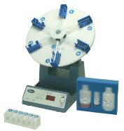
TCS Water Sciences have now developed an automated IMS system, which uses an IMS test developed by ImmuCell Corporation, USA.
The Isolate™ system not only eliminates the RSI risk associated with manual rocking, but also improves the repeatability of the IMS test by standardising the rocking action.
A further advantage of the Isolate™ system is its ability to handle packed pellets up to 2ml, significantly reducing the need for split samples and multiple microscope slides.
In a study comparing the Isolate™ system with a manual IMS test, samples from two sources, a treated upland water and a river water, were filtered and concentrated to give known packed pellet volumes. 10 ml aliquots of each concentrate were spiked with 100 flow-sorted Cryptosporidium oocysts and processed by IMS. Slides were stained and counted, and the counts were expressed as percentage recoveries.
Recovery of Cryptosporidium was significantly better using the Isolate™ system, as seen in Tables 1 and 2. Isolate™ yielded more consistent results than the manual test, as evidenced by the narrower data ranges obtained. These results suggest that the manual rocking procedure is sensitive to variations in operator technique. This study also demonstrated that adjustments to the speed, angle and vigour of the automated rocking procedure had significant effects upon Cryptosporidium recoveries. Once these factors were optimised, consistency of recovery was enhanced.
Table 1: Percentage Recovery of Cryptosporidium from an Upland Treated Water | Manual IMS | Manual IMS | Isolate™ | Isolate™ | | Packed Pellet | 0.5ml | 1.0ml | 0.5ml | 1ml | | Mean (n=10) | 49.7 | 48.4 | 83.7 | 80.4 | | Range | 30-82 | 72-92 | 72-92 | 62-89 |
Table 2: Percentage Recovery of Cryptosporidium from a River Water | Packed Pellet | Manual IMS | Manual IMS | Isolate™ | Isolate™ | | Mean (n=10) | 54.2 | 30.0 | 68.1 | 75.4 | | Range | 45-66 | 25-41 | 60-85 | 58-87 | Conclusions
The regulation of Cryptosporidium testing has been a success, resulting in improved laboratory performance and better standardisation of methodologies. The DWI is now implementing changes to encourage laboratories to evaluate new technology. By doing this, laboratories can expect to benefit from improvements in health & safety, working efficiencies and turnaround times. Automation has improved several aspects of the Cryptosporidium method, including filter washing, sample concentration and detection. Now, by applying automation to IMS, further improvements to both standardisation and Cryptosporidium recovery have been achieved.
|